
Introduction to IMPC Embryo Data
Up to one third of homozygous knockout lines are lethal, which means no homozygous mice or less than expected are observed past the weaning stage (IMPC Viability Primary Screen procedure). Early death may occur during embryonic development or soon after birth, during the pre-weaning stage. For this reason, the IMPC established a systematic embryonic phenotyping pipeline to morphologically evaluate mutant embryos to ascertain the primary perturbations that cause early death and thus gain insight into gene function.
As determined in IMPReSS (see interactive diagram here), all embryonic lethal lines undergo gross morphology assessment at E12.5 (embryonic day 12.5) to determine whether defects occur earlier or later during embryonic development. A comprehensive imaging platform is then used to assess dysmorphology. Embryo gross morphology, as well as 2D and 3D imaging are actively being implemented by the IMPC for lethal lines.
Read more in our paper on High-throughput discovery of novel developmental phenotypes, Nature 2016.
Accessing Embryo Phenotype Data
Embryo phenotype data can be accessed in multiple ways:
- Embryo Images: interactive heatmap A compilation of all our Embryo Images, organised by gene and life stage, with access to the Interactive Embryo Viewer, where you can compare mutants and wild types side by side and rotate 2D and 3D images; we also provide access to our external partners' embryo images.
- Embryo Vignettes Showcase of best embryo images with detailed explanations.
- From the FTP site, latest release All our results. Reports need to be filtered by a dedicated column, Life Stage (E9.5, E12.5, E15.5 and E18.5). Please check the README file or see documentation here.
- Using the REST API (see documentation here)
Determining Lethal Lines
The IMPC assesses each gene knockout line for viability (Viability Primary Screen IMPC_VIA_001). In this procedure, the proportion of homozygous pups is determined soon after birth, during the preweaning stage, in litters produced from mating heterozygous animals. A line is declared lethal if no homozygous pups for the null allele are detected at weaning age, and subviable if pups homozygous for the null allele constitute less than 12.5% of the litter.
Lethal strains are further phenotyped in the embryonic phenotyping pipeline. For embryonic lethal and subviable strains, heterozygotes are phenotyped in the IMPC adult phenotyping pipeline.
IMPC Embryo Phenotyping - Goals and Procedures
With up to one third of knockout strains being embryonic lethal, a systematic unbaised phenotyping pipeline was established to perform morphologic and imaging evaluation of mutant embryos to define the primary perturbations that cause their death. From this important insights are gained into gene function.
IMPC centers funded by the NIH Common fund mechanism are delivering the following for All Lines:
- Viability
- Heterozygote E12.5 Embryonic LacZ staining ( 2 mutant animals, wt reference images)
For All Embryonic Lethal Lines, gross morphology is assessed at E12.5 to determine if defects occur earlier or later in development. A comprehensive imaging platform is then used to assess dysmorphology at the most appropriate stage:
| Procedure | Number | Note |
|---|---|---|
| E9.5 Gross morphology | at least 2 homs,2 wt | images optional |
| E9.5 OPT screening | at least 2 homs | reconstructions available |
| E14.5-E15.5 Gross morphology | at least 2 homs, 2 wt | images optional |
| E14.5-E15.5 microCT screening | at least 2 homs | reconstructions available |
| E14.5 HREM | at least 3 homs, 1wt | reconstructions available |
| E18.5 Gross morphology | at least 2 homs | images optional |
| E18.5 microCT | at least 2 homs, 2 wt | reconstructions available |
In addition, the NIH is supporting in-depth phenotyping of embryonic lethal lines with three current awardees.
Trevor William, University of Colorado School of Medicine
Jesse Mager, University of Massachusetts Amherst
2D Imaging
Embryo LacZ

The majority of IMPC knockout strains replace a critical protein coding exon with a LacZ gene expression reporter element. Heterozygote E12.5 embryos from IMPC strains are treated to determine in situ expression of the targeted gene.
See all genes with embryo LacZ images.
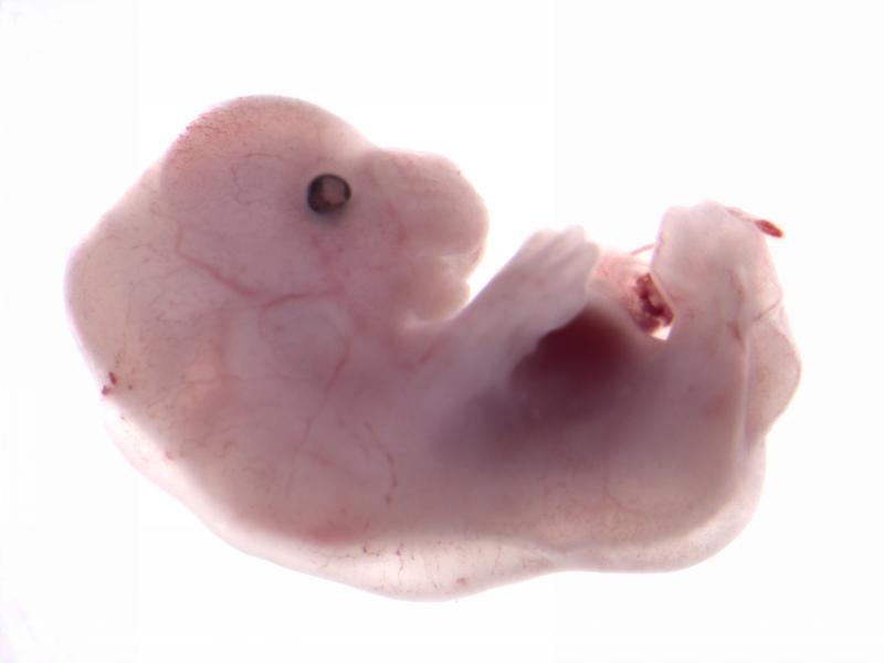
Embryo Gross Morphology
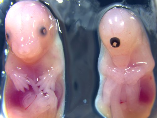
WT / Acvr2a
Gross morphology of embryos from lethal and subviable strains highlights which biological systems are impacted when the function of a gene is turned off. The developmental stage selected is determined by an initial assessment.
See embryo gross morphology images for E12.5, E14.5-E15.5, E18.5.
3D Imaging
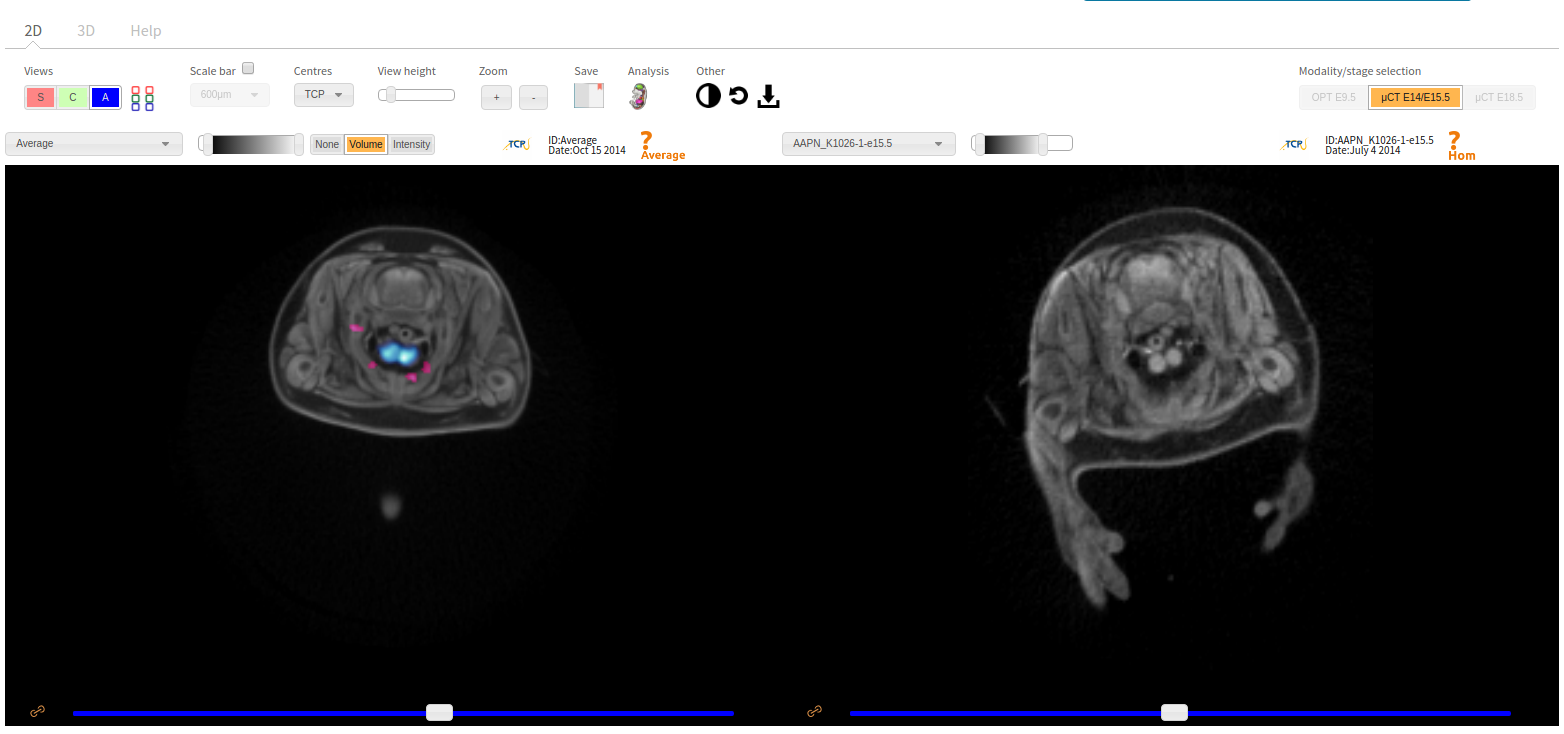
The embryonic and perinatal lethal pipeline comprises several 3D imaging modalities to quantify aberrant morphology that could not be determined by gross inspection. Images acquired by micro-CT and OPT are available via our Interactive Embryo Viewer (IEV).
Vignettes
Chtop has been shown to recruit the histone-methylating methylosome to genomic regions containing 5-Hydroxymethylcytosine, thus affecting gene expression. Chtop mutants showed complete preweaning lethality with no homozygous pups observed. High resolution episcopic microscopy (HREM) imaging, revealed decreased number of vertebrae, abnormal joint morphology and edema. Full Analysis

The Kldhc2 gene is located within a locus linked to an automsomal dominant disease that leads to fibro-fatty replacement of right ventricle myocardium leading to arrythmias (ARVD3 ; OMIM) Full Analysis
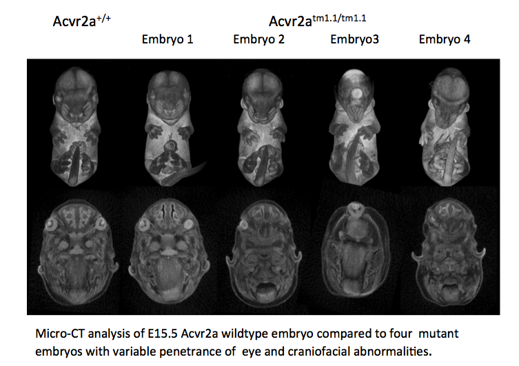
Activin receptor IIA is a receptor for activins, which are members of the TGF-beta superfamily involved in diverse biological processes. Acvr2a mutants are subviable with most pups dying before postnatal day 7. Full Analysis

Chromobox 4 is in the polycomb protein family that are key regulators of transcription and is reported to be upregulated in lung bud formation and required for thymus development Full Analysis
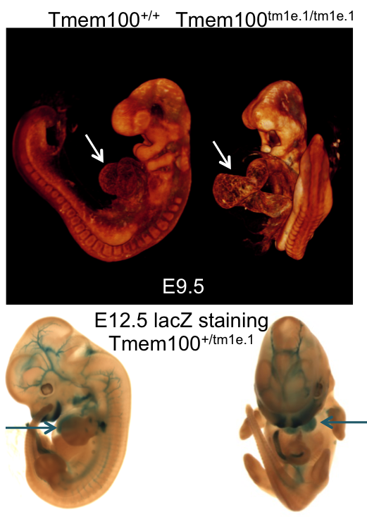
Transmembrane Protein 100 functions downstream of the BMP/ALK1 signaling pathway. Tmem100 mutants showed complete preweaning lethality and were also lethal at E12.5. Full Analysis
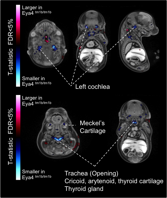
Eyes absent transcriptional coactivator and phosphatase 4 is associated with a variety of developmental defects including hearing loss. Eya4 mutants showed complete preweaning lethality with no homozygous pups observed. Full Analysis
Tox High Mobility Group Box Family Member 3 is a member of the HMG-box family involved in bending and unwinding DNA. Tox3 mutants have partial preweaning lethality with 1/3 of the pups dying before P7. Full Analysis

Radial spoke head protein 9 is a component of the radial spoke head in motile cilia and flagella. Rsph9 mutants showed partial pre-weaning lethality but viable to P7. Full Analysis

Pax 7 is a nuclear transcription factor with DNA-binding activity via its paired domain. It is involved in specification of the neural crest and is an upstream regulator of myogenesis during post-natal growth and muscle regeneration in the adult. Full Analysis
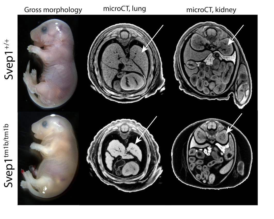
Svep1 codes for an uncharacterized protein named after the multiple, extra-cellular domains identified in the sequence. Homozygotes fail between E18.5 and birth. Full Analysis

Striatins act as both calcium-dependent signaling proteins and scaffolding proteins, linking calcium-sensing signaling events with cellular action. Lethality in Strn3 homozygotes occurs around E15.5. Full Analysis

Rab34 is a member of the RAS oncogene family, involved in intracellular vesicle transport. Rab34 homozygotes are subviable at E18.5. Full Analysis

Cytochrome c oxidase subunit VIIc (Cox7c) is a nuclear-encoded regulatory component of cytochrome c oxidase. Homozygous mutants do not survive between E15.5 and E18.5. Full Analysis
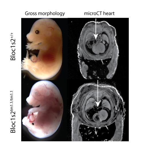
Bloc1s2 functions in the formation of lysosome-related organelles through the BLOC-1 complex, with lethality occurring in knockouts around E15.5. Full Analysis
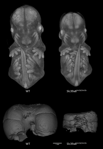
Solute carrier (metal ion transporter) family 39 member 8 (Slc39a8) mutants are small at E14.5 and show heart defects. Full Analysis

Lysine (K)-specific demethylase 8 (Kdm8) is predicted to have dual functions as a histone demethylase and as a protein hydroxylase and is also known as Jmjd5. Tm1b mutants show developmental delay and fail to turn at E9.5. Full Analysis

Autophagy related 3 (Atg3) mutants show cardio-vascular defects at E14.5. Full Analysis
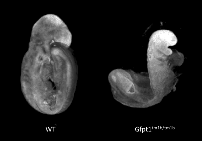
Glutamine:fructose-6-phosphate amidotransferase 1 (Gfpt1) mutants show developmental delay and fail to turn at E9.5. Full Analysis
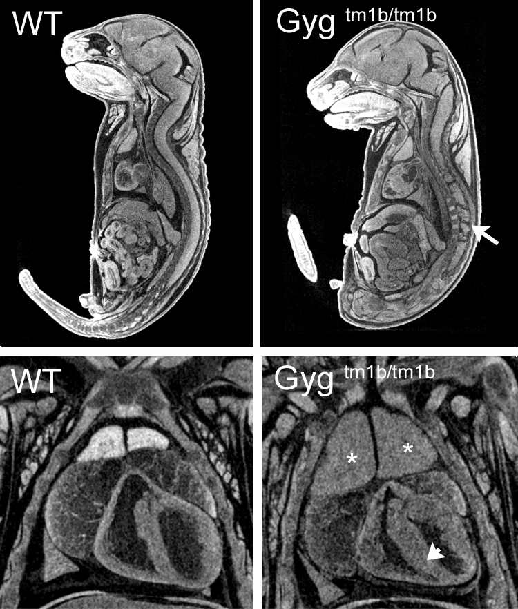
Glycogenin is an enzyme that converts glucose to glycogen. Glycogenin catalyzes UDP-alpha-D-glucose + glycogenin ⇌ UDP + alpha-D-glucosylglycogenin. The enzyme is a homodimer of 37 kDa subunits. Mutations in human GYG1 are associated with Glyocgen Storage Disease XV and Polyglucosan Body Myopathy 2 (OMIM). Homozygous null Gyg mice die between birth and weaning but were found in normal proportions at E18.5. Mutants were indistinguishable from littermates at E12.5, E15.5 or E18.5 but analysis of microCT images revealed obvious cardiac abnormalities, enlarged thymus and abnormal nervous system morphology. This is the first reported Gyg mouse mutant. Full Analysis

Transmembrane protein132a is transmembrane protein of unknown function. Homozygous null mutants were viable at normal proportions at E15.5 and E18.5 but showed obvious and severe defects that were readibly visible by eye. Embryos had abnormal limb morphology with syndactyly, spina bifida, heart abnormalities. Some mutants were smaller than littermates. Full Analysis
These vignettes highlight the utility of embryo phenotyping pipeline and demonstrate how gross morphology, embryonic lacz expression, and high resolution 3D imaging provide insights into developmental biology. Clicking on an image will provide more information.
IMPC Embryonic Pipeline
